Seeing Possibility
A researcher and his team at Stark Neurosciences Research Institute are exploring ways to help the optic nerve connect a donor eye to the brain.
Matthew Harris May 27, 2025
VISION LOSS OFTEN starts with a wiring issue. Strain on the optic nerve compresses delicate cells that transmit visual information from the eye to various processing regions in the brain. Steadily, the connection frays and vision dims. And for some people, it goes dark.
Catching these conditions early is vital, but many treatments only slow deterioration. They cannot restore what has been lost. But what if we could?
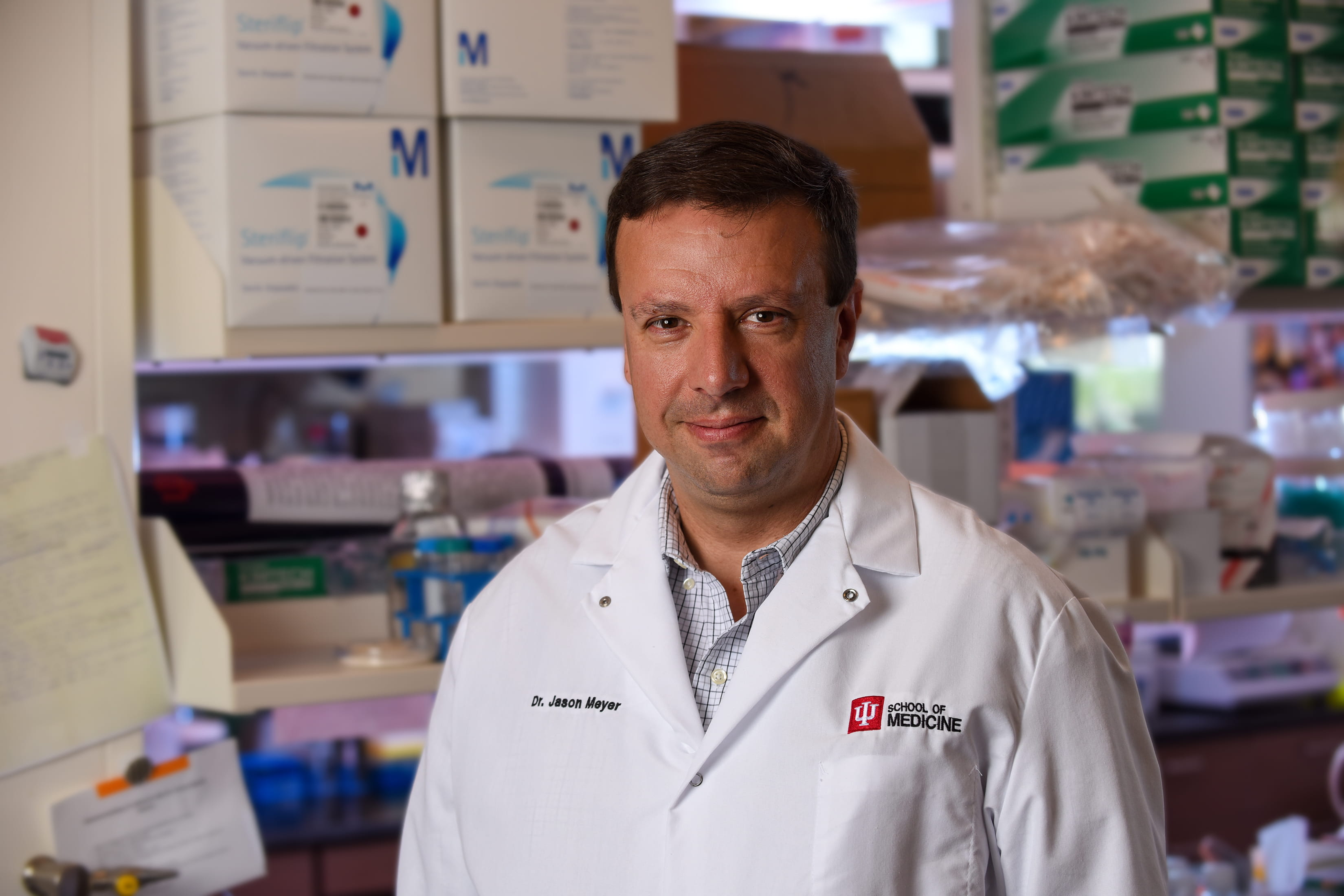
Jason Meyer, PhD, the A. Donald Merritt Professor of Medical & Molecular Genetics at Indiana University School of Medicine, is part of a consortium exploring the feasibility of a seemingly radical solution: whole-eye transplantation. In late December, his lab at the Stark Neurosciences Research Institute earned $2.2 million in funding from the Advanced Research Projects Agency for Health to figure out how stem cells can stimulate the optic nerve to connect with a donor eye.
The breakthrough might be felt most acutely in glaucoma, which afflicts 4.2 million people in the U.S. But once you fold in patients with macular degeneration, diabetic retinopathy and cataracts, up to 54 million Americans with vision-robbing diseases might have a long-term solution.
“If we’re successful, it’s a complete game-changer,” Meyer said. “Whole eye transplantation puts every disease in play. You’re not targeting one specific problem. The approach is the same. Something has gone wrong in a patient’s eye, and we can replace it with a good one.”
While the work is in its infancy, Meyer — and collaborators from the University of Wisconsin and National Eye Institute — are tackling a fundamental problem. Axons are long fibers extending from the end of retinal ganglion cells. They are tiny, only micrometers wide, but must grow several inches to reach the brain.
Even if they complete that trek, it’s all for naught if they don’t tie in at the right junction. Some nerve cells help detect motion. Others distinguish colors by converting visual data from rods and cones. Or they enhance contrasts to detect edges, which is crucial for forming shapes and patterns. That processing work happens in different synapses.
“We’re looking at factors that really enhance the way neurons connect with each other,” Meyer said. “Once they get to the proper point, if they’re not making the right connections, there’s no real success.”
Working together, the team aspires to create a new junction box that a surgeon would install with a transplanted eye. Stored inside would be stem cells, growth factors to spur regeneration, and glial cells that repair and maintain axons. “We’re trying to figure out what kinds of glial cells are best and what kinds of factors they should be producing,” Meyer added.
Meyer’s lab focuses on the biology powering that process. Over the past decade, it has steadily developed a reliable model instrumental in the process. “We had the technical approaches in hand,” he said. “It’s just repurposing them for a new set of questions.”
Meyer started his career interested in neuroscience, initially exploring how motor neurons in the spinal cord are affected by ALS. That work entailed pushing stem cells to become neurons, which could be transplanted and halt a disease that results in paralysis, respiratory failure and, ultimately, death.
Yet Meyer saw that the stem cells he used naturally adopted a profile of cells found in the front of the brain, which are fundamental traits of retinal cells. Over time, Meyer became interested in creating a model that mimicked conditions that caused damage to the optic nerve. The approach differed from how he was taught in grad school, when his mentors worked with stem cells in a flat dish, slathered them in growth factors, and hoped to reap enough mature cells.
The stem cells were already inclined to become retinal cells, and if they were packed together at high enough densities, they started forming a three-dimensional structure. “One of the most important things is not trying to tell cells what to do,” Meyer said. “It’s observing what they’re trying to tell you in order to behave a certain way.”
In the past few years, Meyer’s lab has steadily refined how to grow 3D organoids, also known as mini retinas, suspended in dishes. They stimulate the cells by adding a protein at a specific time, concentration and duration. In doing so, the lab reported that it can efficiently grow mini retinas that are consistently the same size and have enough retinal cells to study.
“We’re hitting them at a point where they know they’re cells in the central nervous system,” Meyer said. “They just haven’t figured out what they want to be when they grow up.”
The consistency of Meyer’s model is its chief allure as part of a consortium — backed by a $46 million ARPH-H award — to make eye transplantation viable. Regenerating aspects of the central nervous system is infinitely more complex than transplanting a liver or kidney. Using his model, Meyer’s lab can conduct dozens of experiments simultaneously to deduce the best strategies to get axons to grow.
And if the group of eight institutions is successful, the possibility of an eye transplant — and a long-term cure — would be in play for millions losing their vision.
“It’s a bold idea,” Meyer said. “There’s very little that can be done (now). This could potentially be something that really helps for the rest of a person’s life.”
To learn how you can support this bold research, contact Sam Kinder, senior director of development, at 317-278-5635 or kindersm@iu.edu